The histopathological laboratory's newly built premises at the new St. Erik Eye Hospital are bright, spacious, ergonomic and well ventilated. Since the move in September 2020, the staff has now had time to establish itself.
"We have designed the rooms and our workflows ourselves and we are content. It has been a period of getting routines to work in the new house, but now everything is starting to settle," says Emma Lardner, biomedical scientist at the histopathological laboratory, St. Erik Eye Hospital.
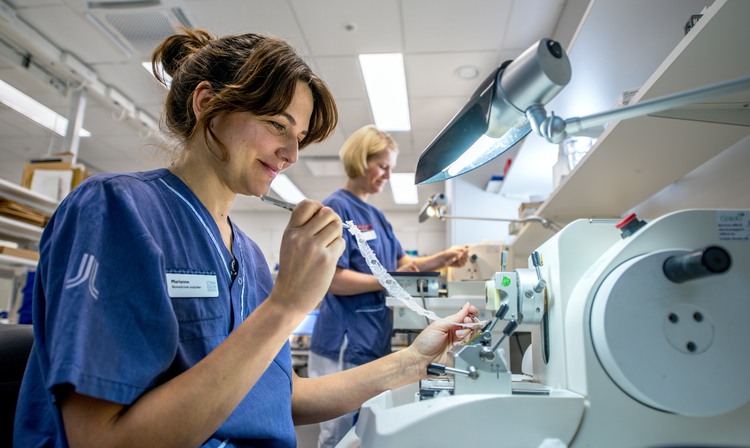
Emma Lardner and Marianne Georgsson handle about 25 tissue samples a week.
Photo: Jens Sølvberg
The histopathological lab receives surgically removed tissue from the eye, eyelids, orbit and lacrimal system, removed in clinics all over Sweden. The most common tissue samples are suspected basal cell carcinoma on and around the eyelids.
Other common diagnoses are squamous cell carcinoma, sebaceous carcinoma, lymphoma, inflammation, vasculitis and actinic keratosis. The biopsies can be millimetre-sized, as in the case of suspected metastases in the eyes. In other cases we receive whole eyes, which is often the case when the eye is affected by uveal melanoma.
However, before the diagnosis can be established by the pathologist, several technical processes are required that need at least two days of preparation. This is where the biomedical scientists Emma Lardner and Marianne Georgsson come into the picture.
.jpg?width=750&height=448&ext=.jpg)
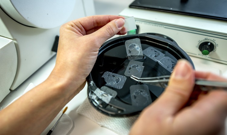
Emma Lardner and Marianne Georgsson take care of the entire process from the sample received to delivery to the pathologists.
Photo: Jens Sølvberg
"Each tissue sample is handled at different stages, both manually and mechanically. If the eye pathologist is to be able to make the correct diagnosis, it is crucial that all parts are optimized," says Marianne Georgsson, biomedical scientist at the histopathological laboratory, St. Erik Eye Hospital.
During the course of a week, she and her colleague Emma Lardner receive an average of 25 tissue samples of varying sizes.
"I like that we get to manage the entire process, from registering the samples received to handing them over to the pathologists. It gives me a strong sense that I am helping the individual patient and am involved in speeding up their diagnosis and treatment," says Emma Lardner.
Extensive process
A regular morning for the biomedical analysts begins with them registering received samples. Then the small biopsies are removed and placed into special boxes, so-called cassettes. The larger tissue samples must be cut into three-millimetre slices.
"It's probably the craftsmanship that I like the most, to stain by hand, cut by hand. It's finicky and tricky and requires a touch of sorts, you have to have your thinking cap on," says Marianne Georgsson.
The tissue samples are embedded in molten paraffin.
Photo: Jens Sølvberg
Once the pieces have been cut out, they are treated in a dehydration machine overnight to allow all the liquid to dissipate. When the water is gone, they are embedded in molten paraffin, a waxy mixture, which is then left to solidify.
"In the morning, the tissue samples are ready. Then, we open the cassettes with pieces of tissue and put each piece into its own mould, then we pour in more paraffin," says Emma Lardner
After that, it is time for the next machine, the microtome, to get to work. The machine cuts each block into four thousandths of a millimetre thin sections. Each small block takes between a few minutes up to half an hour to cut. The thin cuts are picked up with a brush and tweezers and placed in water baths, where they are then fished up with a slide.
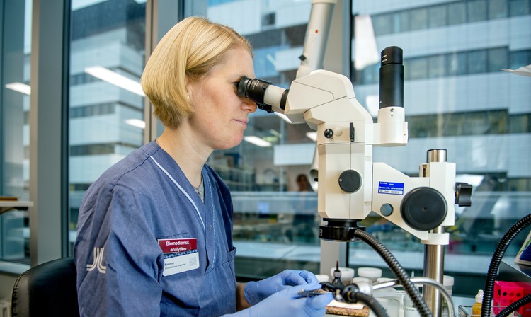
"One cell is ten to twelve thousandths of a millimetre, so our incisions mean that each cell is also divided a few times, which is important in cancer diagnosis. Then you can see the cell nuclei inside and how the cells interact with the rest of the tissue," says Emma Lardner, who herself is very fond of looking at the cells under a microscope, something she always does to assure quality.
Marianne Georgsson likes the craftmanship the most and that the work requires you to really think.
Photo: Jens Sølvberg
Before the tissue samples are finally passed on to the pathologists, the tissue is stained. In routinely used types of stains, the nuclei are coloured blue while the rest of the tissue is made different shades of pink. This way, the pathologist can look at the tissue under the microscope and assess both the appearance of individual cells and structural changes to growth patterns.
"The last thing I do before I go home for the day is to make sure that the stainings look good, then I put everything on a tray and go in to the pathologist," says Marianne Georgsson.
Text: Jenny Ryltenius, Helena Mayer